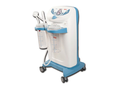
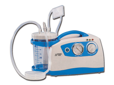
Mediworld Subscription
Subscribe to Mediworld for product discounts, healthcare news, event updates and more!
Bestsellers
The Mediworld product range is extensive, so we have the most popular items for you below.

Load More
Nothing matches the criteria
Mediworld has been a regular supplier to the NHS for a number of decades.
We specialise in turnkey projects and are proudly UN approved.
2023 is our 50 year anniversary, and we are here to service you for many more.
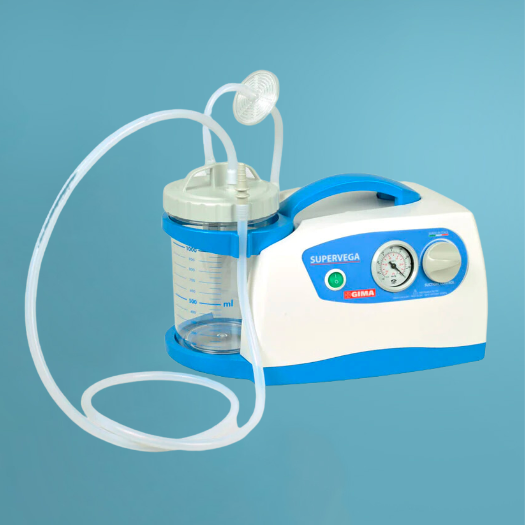
The products that we have are industry leaders for both GP’s & wards.
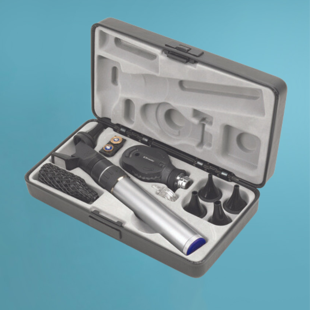
We buy directly from Keeler. Keeler are a major brand within the ophthalmology industry, their specialist diagnostic products, diagnostic sets, and indirect ophthalmoscope, are tools of the trade for many ophthalmic specialists.
ENT simply means Ear, Nose and Throat. A patient would need to get an ENT if they have ear disorders like hearing impairment, ear infections, tinnitus (ringing in the ears), or any form of pain in the ear. ENT specialists can also treat congenital disorders of the ear; these are disorders people are born with.
Both the retinoscope and the ophthalmoscope allow observation of the fundus and the red reflex.
In retinoscopy, there is a required effective light source that is quickly moved off the visual axis. In ophthalmoscopy, this type of illumination is unable to be produced and that’s the major difference between them.
There are 3 different ways ophthalmoscopy is done
• Direct ophthalmoscopy
The patient is seated in a darkened room and the ophthalmologist performs this exam by shining a beam of light through the pupil using the ophthalmoscope.
• Indirect ophthalmoscopy
The patient is expected to either lie or sit in a semi-reclined position, while the ophthalmologist holds the eye open and shines a bright light using a device worn on the head.
Some pressure may be applied to the eye through a blunt probe and it’s usually done to look for a detached retina.
• Slit-lamp ophthalmoscopy
The patient is expected to sit in a chair with the instrument placed in front, and the chin and forehead of the patient are kept on a support to keep the head steady. The ophthalmologist will use the microscope part of the device and the lens placed close to the eye to see. This technique sees in more magnification than indirect ophthalmoscopy.
An otoscope also called an auriscope is a medical device used to look into the ears. It gives a view of the ear canal and eardrum to screen for any ear issues or to investigate ear symptoms
There are 4 different types of filters while using an ophthalmoscope, regardless of the filter's colour, when using the binocular indirect ophthalmoscope, you should use as little light as possible and as much light as required
• No Filter or White Light
This is the light used as a standard light with its true colours at the start of the examination to get an overview of the test.
• Red-free filter (green colour)
This filter produces longer wavelengths and the retina shows a red hue when it’s examined with white light. This allows a clear contrast between the surrounding tissues and the retina and then makes it red-free.
• Yellow filter
The yellow filter is described as a comfort filter for the patient because it reduces photophobia with its warm, soft light. It’s mostly used on sensitive patients and it reduces UV exposures
• Blue filter
This filter is used for differentiating between different layers and membranes of the eye and it’s rarely used by much medical personnel.
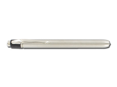
A pen torch alongside a pupil gauge is used to assist with examinations of the eyes, ears or mouth. Although it’s primarily used for the eyes.
Mediworld Subscription
Subscribe to Mediworld for product discounts, healthcare news, event updates and more!
Would you like to save product on your wishlist?
Subscription completed successfully. The letter has been sent by mail.
User already subscribed
