


Blogs
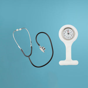
Trust advanced stethoscopes for precise diagnostics and superior patient care.

Different Parts Of The Stethoscope And Their Functions
29.08.2024
What is a stethoscope?
Stethoscopes are essential pieces of medical equipment for any medical professional. It plays a vital function in patient care because medical personnel use a stethoscope to listen to noises generated in the body. Additionally, it can be utilized to detect blood vessel or bowel noises. Hence, this tool is a continuous companion of physicians, nurses, first responders, veterinarians, and other healthcare workers, enabling them to examine heart, lung, and other body noises to detect potential health issues.
The 8 Parts of the Stethoscope and their Functions
Chest Piece
The circular chest piece, commonly known as the head, is the core component of the device. This instrument's part is put against a patient's body. This is the most vital component of a stethoscope. It is designed for detecting, capturing, and transmitting sounds to the headset. The optimal placement of the chest piece is directly against the patient's skin. This consists of the Diaphragm, Bell, and Stem. Also, depending on the model, the chest piece of a stethoscope may be one-sided or two-sided.
Diaphragm
The diaphragm is the spherical and circular end of the chest piece. Its size allows you to examine a more significant portion of the patient's body. It can detect higher-frequency noises throughout a patient. Modern diaphragms reduce cold sensation when placed directly on the patient's skin. This is accomplished by surrounding the diaphragm with a chill ring, which creates an airtight seal and prevents the patient from experiencing the coolness of the portion.
Bell
The bell is the spherical end of the chest piece. It can detect lower-frequency noises than the diaphragm due to its smaller size, making it excellent for smaller body parts. A non-chill ring is also present on the bell's edges.
Tubing
The stethoscope tubing is the flexible rubber or PVC pipe that links the chest piece to the headset. This component of the stethoscope transfers sound waves captured by the diaphragm and bell to the headset with minimal quality loss (which depends on the type of tubing it is built with).
Stem
The stem is the portion of the stethoscope that links the chest piece to the tubing. It comprises metal or steel and guarantees a secure connection for optimum performance. It also permits switching between the diaphragm and the bell.
Headset
The stethoscope's top portion is known as the headset. It is constructed with two ear tubes, tension springs, and ear tips. It facilitates an effective passage of sound into the ear canal so that disturbances are minimized.
Ear Tubes
The ear tubes are hollow metal tubes that link the acoustic tubes to the ear tips. They serve as the left and proper channels for sound transmission. The tension spring lies between the ear tubes, allowing you to regulate the tightness with which the stethoscope fits in your ears.
Ear Tips
The components that enter the ear canal are the ear tips, which are composed of rubber or silicone. They are put on the ends of the ear tubes and feature a hole to allow sound to escape. The ear tips must be soft and stiff to produce a seal in the ear canal and block out all external noise.
Different Types of Stethoscopes
Acoustic Stethoscope
The acoustic stethoscope functions by directing more sound waves to the ear than would usually reach it. For humans to hear a sound, sound waves must force air molecules to vibrate, resulting in changes in air pressure that cause our eardrums to vibrate. When a stethoscope is placed on a patient, sound waves generated by bodily noises, such as a heartbeat or gurgling stomach, strike the metal chest piece of the stethoscope.
The rubber tube then directs the sound waves until they reach the metal earpiece and, subsequently, your ears. Because the tube contains the sound waves, more of them reach your ears than would otherwise be the case, enhancing the sound. This is why using a stethoscope to listen to a patient's heart sounds louder than placing your ear directly on their chest.
Electronic stethoscopes
Electronic stethoscopes (digital stethoscopes) convert the sound's physical vibrations into an electronic signal and optimize them for enhanced listening and diagnosis. In addition to the capacity to increase the loudness, many electronic stethoscopes may also minimize background noise. Some electronic stethoscope types are constructed as a chest piece that connects directly to a set of headphones. The majority of electronic stethoscope devices resemble traditional stethoscopes. Electronic stethoscopes are either battery-powered or rechargeable; therefore, if the battery is dead or you forget to charge it, the device will not function.
Stethoscopes for Hearing Impaired
There are various choices accessible for medical professionals with hearing impairments. As explained in the section just above, an amplified electronic stethoscope will raise the level of the sounds, making them more audible. This stethoscope can be used generally if you do not wear hearing aids. If you wear hearing aids, you can experiment with special adaptors known as stethoscope tips that allow you to use a stethoscope with your hearing aids.
If wearing a conventional stethoscope with hearing aids is bothersome, you can also use a digital stethoscope into which headphones can be plugged. As long as you select sufficiently large headphones (consider over-ear and on-ear varieties), they should fit nicely over your hearing aids. Depending on the model of your hearing aids and digital stethoscope, you may be able to acquire a transmitter that wirelessly transmits sounds from the stethoscope to the hearing aids.
Conclusion
Compared to other medical devices, the stethoscope appears to be relatively small. However, when assessing the stethoscope's components, these instruments are designed to be one of the most used diagnostic tools used by medical professionals in determining a clinical condition by listening to the stomach and vessels.
FAQ
How does a Stethoscope detect sound?
The stethoscope's disc and tube amplify and make louder small sounds that are made by the heart, lungs, and other organs of the patient. The doctor listens through the earpieces as the enhanced sounds ascend the stethoscope's tube.
Why are there 2 sides to a stethoscope?
The bell and the diaphragm are the two separate sound-receiving heads on the stethoscope. The diaphragm is used to detect high-frequency sounds, while the bell is used to detect low-frequency noises
Been wanting to buy your own Stethoscope? Visit Mediworld to shop for the best Stethoscopes.
Learn More Now

How to use a Stethoscope: A Beginners Ultimate Guide
12.10.2023
The heart, lungs, and intestines all make sounds that can be heard with a stethoscope. Auscultation is the process of hearing sounds with a stethoscope. Stethoscopes are used by people who are trained to use them, but anyone can learn how to use one. Learn how to use a stethoscope by reading on.1. How do you use a stethoscope
Get a high-quality stethoscope
It is important to have a good stethoscope. The better your stethoscope is, the easier it will be to listen to what's going on inside your patient's body.
Stethoscopes with one tube are better than ones with two. With double-tubed stethoscopes, the tubes can rub against each other. Because of this noise, it may be hard to hear clearly .
If you don't want to wear the stethoscope around your neck, the best tubing is thick, short, and stiff. In that case, you should use a longer tube.
Tap on the diaphragm (the flat side of the chest piece) to make sure there are no leaks in the tube. Use the ear pieces to listen for sounds as you tap. There may be a leak if you don't hear anything.
Adjust your stethoscope’s earpieces
It's important to make sure that the earpieces fit well. If you don't, your stethoscope might not let you hear anything.
Make sure the earpieces are facing forward. You won't be able to hear anything if you put them in backwards.
Make sure the earpieces fit well and have a good seal to keep out noise from the outside.
Most stethoscopes have ear pieces that can be taken off if they don't fit well. Go to a store that sells medical supplies to buy different earpieces.
Some stethoscopes let you tilt the earpieces forward to make sure they fit better.
Check the earpiece tension on your stethoscope
In other words, make sure the earpieces are close, but not too close, to your head. Adjust your earpieces if they are too tight or too loose.
If the earpieces aren't tight enough, you might not hear anything. To make the tension tighter, squeeze the headset where the earpieces are.
If the earpieces are too tight, they might hurt your ears and make it hard to use the stethoscope. Pull the headset apart gently to ease the tension.
Choose an appropriate chest piece for your stethoscope
There are a lot of different types of stethoscope chest pieces. Choose the one that best fits your needs. There are different sizes of chest pieces for adults and children.
2. Listening to the heart
Hold the diaphragm over the patient’s heart
Place the diaphragm on the left side of the upper chest, where the fourth and sixth ribs meet.This is almost right under the breast. Hold the stethoscope between your pointer and middle fingers and apply just enough gentle pressure so that you can't hear your fingers rubbing together.
Listen to the heart for a full minute
Ask the person to calm down and take normal breaths. You should hear the normal sounds of the heart, which sound like "lub-dub." You can also call these sounds "systolic" and "diastolic." The systolic sound is "lub," and the diastolic sound is "dub."
When the mitral and tricuspid valves of the heart close, they make the "lub" or "systolic" sound.
The "dub" sound, called diastolic, comes from the closing of the aortic and pulmonic valves.
Count the number of heartbeats you hear in a minute
Adults and children over 10 years old have a normal resting heart rate of between 60 and 100 beats per minute. For well-trained athletes, the normal resting heart rate may only be between 40 and 60 beats per minute.For people under 10 years old, there are several different ranges of resting heart rates to think about. Among these ranges are:
70–190 beats per minute for babies up to one month old
80–160 beats per minute for babies 1–11 months old
80–130 beats per minute for children 1–2 years old
80–120 beats per minute for children ages 3–4.
Children ages 5 to 6: between 75 and 115 beats per minute
7–9-year-old children: 70–110 beats per minute
Listen for abnormal heart sounds
You should also listen for any strange sounds as you count the heartbeats. Anything that doesn't sound like "lub-dub" might be thought of as strange. If you hear anything strange, a doctor may need to look at your patient again.
If you hear a "lub...shhh...dub" sound or a "whooshing" sound, your patient may have a heart murmur. When blood moves quickly through the valves, this is called a heart murmur. People often have heart murmurs that aren't dangerous. But some heart murmurs can be signs of problems with the heart valves. If you hear a heart murmur, you should tell your patient to see a doctor.
If you hear a third heart sound that sounds like a low-frequency vibration, your patient might have a ventricular defect. This third heart sound is called a ventricular gallop or S3. If you hear a third heart sound, tell the person to see a doctor.
You can find out if what you are hearing is normal by listening to recordings of normal and abnormal heart sounds.
3. Listening to the Lungs
Ask your patient to sit straight up and breathe normally
As you listen, you can ask the patient to take a deep breath if you can't hear their breath sounds or if they are too quiet to tell if anything is wrong.
Use the diaphragm of your stethoscope to listen to your patient’s lungs
Listen to the upper and lower lobes of the patient's lungs, as well as the front and back of the patient's lungs.
As you listen, put the stethoscope on the top of the chest, then the middle of the clavicle, and finally the bottom of the chest. Listen to both the front and back of each of these areas.
Make sure to look at both sides of your patient's lungs and write down anything that seems out of the ordinary.
By using all of these positions, you will be able to listen to all of the lobes of your patient's lungs.
Listen for normal breathing sounds
Sounds of normal breath are clear, like when someone blows into a cup. Listen to a sample of healthy lungs and then compare what you hear to what you hear in your patient's lungs.Normal breath sounds come in two forms:
Breath sounds heard in the tracheobronchial tree are called "bronchial."
Sounds made by breathing that are heard over the lung tissue are called vesicular.
Listen for abnormal breath soundsWheezing, stridor, rhonchi, and rales are all abnormal breath sounds. If you don't hear any breath sounds, it could mean that there is air or fluid around the lungs, that the chest wall is thick, or that the airflow to the lungs is either too slow or too fast.There are four kinds of breath sounds that shouldn't be there:
When a person who wheezes breathes out, or sometimes when they breathe in, they make a high-pitched sound. Many asthma patients also wheeze, and sometimes you can hear the wheezing even without a stethoscope.
Most of the time, when a person with stridor breathes in, it sounds like high-pitched musical breathing that sounds like wheezing. A blockage at the back of the throat causes stridor. Most of the time, you can hear this sound even without a stethoscope.
The sound of Rhonchi is like snoring. Rhonchi is a sound that can't be heard without a stethoscope. It happens when air moves through the lungs in a "rough" way or is blocked.
Rales sounds like bubble wrap popping or something shaking in the lungs. When a person breathes in, rales can be heard.
4. Listening to Abdominal Sounds
Place the diaphragm on your patient’s bare stomach
Use the belly button of your patient as a center point and divide your listening into four parts around the belly button. Listen to the left, right, bottom left, and bottom right.
Listen for normal bowel sounds
Normal bowel sounds sound like a growling or rumbling stomach. Anything else could mean that something is wrong and the patient needs to be looked at more closely.
In each of the four parts, you should hear "growling." Sometimes, it takes a while for bowel sounds to come back after surgery.
Listen for abnormal bowel soundsWhen you listen to your patient's bowels, most of the sounds you hear are just the sounds of digestion. Most bowel sounds are normal, but anything out of the ordinary could be a sign of a problem.If you aren't sure if the sounds you hear coming from the patient's bowels are normal or if they have other symptoms, they should see a doctor to be checked out.
If you don't hear any bowel sounds, it could mean that the patient's stomach is blocked. It can also be a sign of constipation, and the sounds of the bowels may come back on their own. But if they don't come back, there may be something in the way. In this case, the patient needs to be checked out by a doctor again.
If the patient has loud bowel sounds followed by no bowel sounds, that could mean that the bowel tissue has been damaged or has died.
If the patient's bowel sounds are very high-pitched, this could mean that the patient's bowels are blocked.
Drugs, spinal anesthesia, infections, injuries, abdominal surgery, or overexpansion of the bowel can all cause slow bowel sounds. Crohn's disease, a gastrointestinal bleed, food allergies, diarrhea, an infection, or ulcerative colitis can all cause bowel sounds that are fast or too loud.
5. Listening For a Bruit
Determine if you need to check for a bruit
If you hear a sound that sounds like a heart murmur, you should also check for a bruit. Since heart murmurs and bruits sound the same, it is important to check for both if one is suspected.
Place the diaphragm of your stethoscope over one of the carotid arteries
The carotid arteries are on either side of the Adam's apple in the front of your patient's neck. If you run your index finger and middle finger down the front of your throat, you can see where your two carotid arteries are.
Be careful not to press too hard on the artery, or you could stop the blood flow and make the person pass out. Don't ever put pressure on both carotid arteries at once.
Listen for bruits
A bruit is a whooshing noise that means an artery is getting smaller. Sometimes a bruit and a murmur are mistaken for each other because they sound the same, but if a patient has a bruit, the whooshing sound will be louder in the carotid artery than in the heart.
You might also want to listen for bruits over the abdominal aorta, renal arteries, iliac arteries, and femoral arteries.
6. Checking Blood Pressure
Wrap the blood pressure cuff around your patient’s arm, right above the elbow
If the patient's sleeve is in the way, roll it up. Use a blood pressure cuff that fits the arm of your patient. You should be able to wrap the cuff around your patient's arm so that it fits snugly but is not too tight. Get a different size cuff if the one you have is too small or too big.
Press the diaphragm of the stethoscope over the brachial artery just below the cuff's edge
If you have trouble hearing with the bell, you can also use the diaphragm. You will be listening for Korotkoff sounds, which are low-pitched knocking noises that show the systolic blood pressure of the patient.
Find your pulse on the inside of your arm to figure out where your brachial artery is.
Inflate the cuff to 180mmHg or 30mm above your expected systolic blood pressure
Look at the sphygmomanometer, which is the gauge on the blood pressure cuff, to find the reading. Then, let air out of the cuff at a slow pace (3mm/sec). As you let out the air, use the stethoscope to listen and keep an eye on the sphygmomanometer (gauge on the blood pressure cuff).
Listen for Korotkoff sounds
The first sound you hear is the systolic blood pressure of your patient. Keep an eye on the sphygmomanometer and remember that number. Note the number that the first sound stops on after it stops. The diastolic pressure is equal to that number.
Release and remove the cuff
As soon as you get the second number, let the blood pressure cuff go down and take it off the patient. When you're done, you should have two numbers that add up to the blood pressure of your patient. Put these numbers next to each other and put a slash between them. 110/70 is an example.
7. Wait a few minutes if you want to check the patient’s blood pressure again
If the patient's blood pressure is high, you might want to take it again
Your patient may have high blood pressure if their systolic blood pressure is over 120 or if their diastolic blood pressure is over 80. If that's the case, your patient should see a doctor for more tests.
FAQsWhat is a heart murmur?
A heart murmur is an extra or strange sound coming from the heart that can be heard with a stethoscope and may be a sign of a problem with the heart.
Are all stethoscopes the same?
No matter what kind of stethoscope you want, whether it is specialized or not, it will have the same basic parts and design.CTA: Doyou want toknow more about this? Mediworld is the perfect placeto view medical equipment and more!
Learn More Now