


Blogs

Overview of the different ways you can use a stethoscope
18.07.2024
An overview on the uses of a Stethoscope
In this section, we will review the many different sounds a health practitioner can look out for when using a stethoscope. Keep in mind that this is a general list that should be used as a guide only.
1. Diagnosing cardiovascular and respiratory conditions
A stethoscope is placed on the chest to listen to the sound of the heart beating and the lungs breathing.
Heart sounds, such as the "lub-dub" of the heart valves closing, can indicate the presence of certain conditions, such as a heart murmur, valve problems, or an irregular heartbeat.
Lung sounds, such as crackles or wheezes, can indicate the presence of conditions such as pneumonia, asthma, or chronic obstructive pulmonary disease (COPD).
2. Assessing blood flow through the arteries in the neck (carotid artery) or legs (peripheral artery disease)
A stethoscope is placed on the neck to listen for the sound of blood flowing through the carotid artery supplies blood to the brain.
By measuring the sound and the timing of the sound, a doctor can assess the blood flow and detect any blockages or narrowing of the artery, indicating an increased risk of stroke.
A stethoscope can also be used to listen to the blood flow in the peripheral artery of the legs, which can help identify peripheral artery disease (PAD)
3. Detecting fluid in the lungs (pleural effusion) or around the heart (pericardial effusion)
A stethoscope is placed on the chest to listen for abnormal sounds that can indicate the presence of fluid in the lungs (pleural effusion) or around the heart (pericardial effusion). The presence of fluid can indicate severe conditions such as heart failure, pneumonia, or cancer.
4. Identifying bowel sounds to assess intestinal motility
A stethoscope is placed on the abdomen to listen for the sounds of the intestines contracting and pushing food through the digestive system.
The presence or absence of bowel sounds, or changes in the normal pattern of bowel sounds, can indicate a problem with intestinal motility and may suggest conditions such as constipation, obstruction, or peritonitis.
5. Evaluating the function of heart valves
A stethoscope is placed on the chest to listen to the sound of the heart valves opening and closing.
By listening to the sound and timing of the valves, a doctor can evaluate the function of the valves and detect any problems, such as leakage (regurgitation) or narrowing (stenosis) of the valves.
6. Detecting murmurs and other unusual sounds that may indicate a problem
A stethoscope is placed on the chest to listen for any murmurs or other unusual sounds that may indicate a problem with the heart or lungs.
Murmurs, for example, can indicate problems such as a heart valve problem or anemia.
7. Assessing lung expansion and breathing patterns in patients with lung disease
A stethoscope is placed on the chest to listen to the sound of the lungs inflating and deflating.
By assessing the sound and pattern of breathing, a doctor can determine if there is a lung problem, such as emphysema or asthma.
8. Performing a diaphragm test to evaluate for diaphragm paralysis
A stethoscope is placed on the chest to listen to the sound of the diaphragm, the muscle that helps a person breathe, moving up and down.
By assessing the sound and movement of the diaphragm, a doctor can determine if there is a problem with the diaphragm, such as paralysis, which can make breathing difficult.
9. Listening to the fetus’s heartbeat during pregnancy
A stethoscope is placed on the mother's abdomen to hear the heartbeat of the fetus’s heartbeat.
By listening to the heart rate, a doctor can assess the fetus’s health and ensure that everything is generally progressing during the pregnancy.
10. Checking for blood pressure by listening for the pulse in the arteries
A stethoscope is placed on the artery, usually the brachial artery in the arm, to listen for the sound of the pulse.
By using a blood pressure cuff to occlude the artery and measuring the timing of the sound, a doctor can calculate the systolic and diastolic blood pressure.
11. Monitoring breathing rate and pattern to diagnose certain conditions
A stethoscope is placed on the chest to listen to the sound of the lungs inflating and deflating and the rate and pattern of breathing.
By assessing the sound and pattern of breathing, a doctor can determine if there is a lung problem, such as asthma or chronic obstructive pulmonary disease (COPD).
12. Checking for abnormal sounds in the abdomen
A stethoscope is placed on the abdomen to listen for any abnormal sounds that may indicate a problem with the organs or digestive system.
Abnormal sounds such as gurgling, high-pitched noises, or absence of bowel sounds can indicate issues such as obstruction, peritonitis, or constipation.
13. Listening for bowel sounds to indicate proper digestion
A stethoscope is placed on the abdomen to listen for the sound of the intestines contracting and pushing food through the digestive system.
The presence or absence of bowel sounds, or changes in the typical pattern of bowel sounds, can indicate a problem with intestinal motility and may suggest conditions such as constipation, obstruction, or peritonitis.
14. Diagnosing ear infections by listening for fluid buildup in the middle ear or other abnormalities in the ear canal
A stethoscope is placed on the ear canal to listen for any fluid buildup in the middle ear or other abnormalities in the ear canal.
The presence of fluid or other abnormalities can indicate an ear infection or other ear condition.
15. Checking fetal heart rate during pregnancy to monitor the baby's health.
A stethoscope is placed on the mother's abdomen to listen to the sound of the fetus' heart beating.
By listening to the heart rate, a doctor can assess the fetus’s health and ensure that everything is generally progressing during the pregnancy.
Learn More Now

Stethoscope 101: Listen to the Lungs
18.07.2024
Stethoscope 101: Listen to the Lungs
The stethoscope is medical equipment that is commonly used by healthcare professionals to listen to the sounds of the body. One of the most important uses of the stethoscope is to listen to the lungs.
When listening to the lungs through a stethoscope, healthcare professionals can hear different sounds that are associated with different symptoms. These sounds can give healthcare professionals important information about the health of the patient.
Step 1: Prepare the patient and the environment
Prepare the environment for optimal listening conditions, and make sure the patient is comfortable (either sitting or standing) in a relaxed position. You can also warm the stethoscope in your hands to avoid discomfort for the patient.
Step 2: Instruct the patient on proper breathing
Ask the patient to breathe slowly and deeply through their mouth to maximise lung expansion, and instruct the patient to avoid talking during auscultation.
Step 3: Systematically work your way down the lungs
Compare the left and the right lung sounds symmetrical by starting at the top of the lungs and working your way downwards while moving from one lung to the other. Examine both the anterior and posterior lung fields.
Step 4: Identify the sounds
One of the most common sounds that healthcare professionals listen for is called the breath sound. This sound is produced by the movement of air through the lungs when a person breathes. The breath sound is usually a soft, rustling noise that can be heard throughout the lungs.
However, if there is a problem with the lungs, the breath sound can change. For example, if there is fluid in the lungs, the breath sound can become crackling or bubbling. This sound is known as the crackling sound and is often heard in patients with pneumonia or congestive heart failure.
Another sound that healthcare professionals listen to is called the wheezing sound. This sound is produced by the narrowing of the airways in the lungs. The wheezing sound is a high-pitched whistling noise that can be heard when a person breathes in or out. This sound is often heard in patients with asthma or chronic obstructive pulmonary disease (COPD).
In addition to the crackle and wheeze sounds, healthcare professionals also listen for the pleural friction rub sound. This sound is produced when the two layers of tissue surrounding the lungs rub together. The pleural friction rub sound is a creaking noise that is often heard in patients with pleurisy or pneumonia.
The different sounds that healthcare professionals listen to can provide valuable information about the health of the patient.
Learn More Now

Stethoscope 101: Listen to the Heart
18.07.2024
Stethoscope 101: Step-by-step guide to Listening to the Heart
Learning how to use a stethoscope to listen to the heart is an essential skill for medical professionals, especially those in the cardiology field. The stethoscope helps to identify and diagnose various heart conditions, including murmurs, valve disorders, and arrhythmias. Here is a step-by-step guide on how to listen to the heart and the common sounds to look out for when using a stethoscope.
Step 1: Positioning the Stethoscope
Before placing the stethoscope on the patient's chest, it's crucial to ensure that it's in good working condition. Check the earpieces and diaphragm for any cracks or damage that may affect the sound quality. Once you're satisfied with the stethoscope's condition, place the earpieces in your ears and position the diaphragm on the patient's bare chest, slightly to the left of the sternum, which is the bone that runs down the centre of the chest. You will also need to select the appropriate stethoscope component and use the diaphragm side to detect higher frequencies.
Step 2: Identifying the Heart Sounds
The heart has four distinct sounds, which are commonly referred to as S1, S2, S3, and S4. S1 is the first sound heard and is caused by the closure of the mitral and tricuspid valves. S2 is the second sound heard and is caused by the closure of the aortic and pulmonary valves. S3 and S4 are the third and fourth sounds, respectively, and are caused by the blood flowing into the ventricles.
Step 3: Listening to the Heart Sounds
First, you will have to identify the auscultation points on the chest:
The aortic valve area is found on the second intercostal space on the right sternal border
The pulmonic valve area is found at the fourth intercostal space on the left sternal border
The mitral valve is at the fifth intercostal space in the midclavicular line.
To listen to the heart sounds, place the diaphragm on the patient's chest and listen for the four sounds. S1 and S2 are the most prominent sounds and are heard in all patients. S3 and S4 are fainter and may only be heard in some patients. It's essential to listen to each sound individually and note any abnormalities or irregularities in its timing or intensity.
Step 4: Identifying Heart Murmurs
Heart murmurs are caused by the turbulent flow of blood through the heart valves. They can be heard as a whooshing or swishing sound between the S1 and S2 sounds. Murmurs can be classified as systolic, diastolic, or continuous, depending on when they occur in the cardiac cycle. It's crucial to note the timing, intensity, and location of the murmur and refer the patient to a cardiologist for further evaluation if necessary.
In conclusion, learning how to use a stethoscope to listen to the heart is an essential skill for medical professionals. By following the steps outlined above, you can identify the four heart sounds and the common murmurs heard in patients. Regular practice and experience will help you become more proficient in identifying and diagnosing various heart conditions.
Step 5: Assess Heart Rate and Rhythm
Count the number of beats per minute (BPM) by listening to the heart for 60 s. You can then determine if the heart beat is regular or irregular and note down any variations in the intensity or timings.
Learn More Now

Stethoscope 101: Analysing sounds in the Bowels
18.07.2024
Stethoscope 101: Analysing sounds in the Bowels
Bowel sounds are the noises that are made by the movement of food, liquids, and gas in the gastrointestinal tract. These sounds can be heard using a stethoscope, a medical device used to listen to internal sounds in the body. Identifying different bowel sounds and their significance can provide valuable information about a patient’s digestive health.
Types of Bowel Sounds
There are several types of bowel sounds that can be heard using a stethoscope. The four main types are:
Normal Bowel Sounds: These are the sounds heard when the digestive system is functioning correctly. Normal bowel sounds are characterized by a series of soft clicks and gurgles that occur irregularly every 5 to 15 seconds.
Hyperactive Bowel Sounds: These are bowel sounds that are louder and more frequent than normal bowel sounds. They can be heard when there is an increase in the movement of the digestive tract. Hyperactive bowel sounds can be a sign of diarrhoea, gastroenteritis, or other digestive disorders.
Hypoactive Bowel Sounds: These are bowel sounds that are abnormally quiet or absent. They can be heard when there is a decrease in the movement of the digestive tract. Hypoactive bowel sounds can be a sign of constipation, abdominal surgery, or other digestive disorders.
Absent Bowel Sounds: These are bowel sounds that are not heard at all. They can be a sign of a serious medical condition, such as bowel obstruction or paralytic ileus.
Significance of Bowel Sounds
By listening to bowel sounds, healthcare professionals can gather important information about a patient’s digestive health. Abnormal bowel sounds can be a sign of various digestive disorders, including:
Irritable bowel syndrome (IBS)
Crohn’s disease
Ulcerative colitis
Diverticulitis
Gastroenteritis
Bowel obstruction
Additionally, bowel sounds can be used to monitor a patient’s recovery after abdominal surgery or to track the progress of a patient’s treatment for a digestive disorder.
In conclusion, identifying different bowel sounds and their significance on a patient’s health is an important skill for healthcare professionals. By using a stethoscope to listen to bowel sounds, healthcare professionals can gather valuable information about a patient’s digestive health and make informed decisions about their treatment.
Learn More Now

Stethoscope 101: Detecting Body Fluids
18.07.2024
Stethoscope 101: Detecting Body Fluids
A stethoscope is a medical device used by healthcare professionals to listen to the sounds produced by the body. It is commonly used to detect heart, lung, and bowel sounds. However, did you know that it can also be used to detect different types of bodily fluids?
Fluid in the Lungs: Pleural Effusion
One of the bodily fluids that can be detected using a stethoscope is pleural effusion. Pleural effusion is the buildup of fluid in the space between the lungs and the chest wall. When a healthcare professional listens to the chest with a stethoscope, they may hear a dull sound, which can indicate the presence of fluid in the lungs.
Fluid build-up in the Abdomen: Ascites
Another bodily fluid that can be detected using a stethoscope is ascites. Ascites is the buildup of fluid in the abdomen. When a healthcare professional listens to the abdomen with a stethoscope, they may hear a shifting or "splashing" sound, which can indicate the presence of fluid in the abdomen.
Fluid in the heart: Pericardial Effusion
Pericardial effusion is another bodily fluid that can be detected using a stethoscope. Pericardial effusion is the buildup of fluid in the sac surrounding the heart. When a healthcare professional listens to the heart with a stethoscope, they may hear a muffled or distant sound, which can indicate the presence of fluid around the heart.
A stethoscope can be a useful tool for detecting different types of bodily fluids. Healthcare professionals can listen to the sounds produced by the body to determine the presence of fluid in the lungs, abdomen, or around the heart. It is important to note that a stethoscope should not be used as the sole method of diagnosis, and other diagnostic tests may be necessary to confirm a diagnosis.
Learn More Now

Stethoscope 101: How to use a Stethoscope
18.07.2024
Stethoscope 101: How to use a Stethoscope
The first thing that springs to mind when you think of the medical field is a white lab coat, a stethoscope, and maybe prescription glasses but even while we are all familiar with the stethoscope, very few of us truly know how to operate this iconic medical equipment.
Using a stethoscope correctly requires practice, knowledge and skill. The nuances of this specialised tool can be tricky to master. It is important that you understand how to use a stethoscope correctly in order to best benefit your patients. Doctors or healthcare professionals will have to learn to accurately and quickly diagnose illnesses in their patients. Practitioners can become excellent at swiftly recognizing normal vs abnormal sounds within different parts of the body when practice and experience are put into play.
This guide will take you through the process of using a stethoscope correctly to ensure accurate diagnoses and monitoring of your patient's health. Let's dive into the world of stethoscopes!
Getting to Know Your Stethoscope
In this section, we will cover the basics of using a stethoscope correctly, from proper placement and pressure on the patient's chest to the positioning of your ears for optimal sound clarity. We will also discuss the different types of sounds you should be listening for and key factors that may affect sound quality. Additionally, we’ll go over some tips and tricks for mastering this valuable tool.
The Parts of a Stethoscope
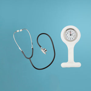
Before we jump into using a stethoscope, it's essential to understand its components and functions. A stethoscope comprises earpieces, tubing, and a chest piece with a diaphragm and bell. Each part plays a crucial role in transmitting internal body sounds to your ears.
A stethoscope has three main parts: the earpieces, the tubing, and the chest piece. The chest piece is made up of a diaphragm, a bell and, in some cases, additional attachments that allow for better sound transmission. The diaphragm is located on one side of the chest piece, while the bell is placed on the opposite side.
The ear tubes are connected to the chest piece, and they go over your ears so that you can hear sounds clearly. These tubes can be adjusted so they fit comfortably – not too tight or too loose – around your ears. Additionally, you may need to adjust the angle in which they lie against your head in order to find the best fit for you.
The diaphragm is placed on the patient to listen to their heart, lungs, and other organs. The bell is placed on the patient first, as it picks up low-pitched sounds such as those made by the heart and lungs during respiration. Meanwhile, the diaphragm can pick up higher-pitched noises like those coming from the intestines, blood vessels, and joints.
Step-by-Step Guide to Using a Stethoscope
Here are the steps you need to take to ensure success:
Preparing the Patient for the Procedure
Introduce yourself. Get the patient comfortable, introduce yourself and explain who you are and why you are using a stethoscope. This can help put the patient at ease and ensure they are relaxed during the procedure.
Check the patient's medical history. The next big thing is inquiring about any medical conditions they may have or have had that can affect the results of your examination.
Stethoscope Positioning
Positioning your stethoscope correctly can make a huge difference in your ability to diagnose a patient.
Here are a few steps to follow to make sure you get the best results in your stethoscope examination:
Place the stethoscope's ear tips in your ears, positioning it snugly so there are no air gaps between them and the outer ear canal.
Place the diaphragm on the patient's skin by lightly pressing it down with your index finger or palm
When using the bell, you should hold it firmly against your patient's skin with two fingers — usually your index and middle finger — while keeping the rest of your fingers away from their body so you don't affect the sound of their heartbeat or breathing.
Finally, keep your hand flat against their body and move it slowly over the designated area to locate sounds that may indicate abnormalities — this will help you distinguish between normal and abnormal sounds.
Listening and Recording Findings
Paying attention to the sounds that you hear through the stethoscope is very important. This is because it helps you accurately determine the diagnosis of your patient. When auscultating, or listening for sounds, take care in distinguishing between subtle variations of sound quality, intensity, and pitch. It is also important to note abnormal sounds such as extra heart sounds, abnormal respiratory noises, and murmurs. It is essential that you take notes to record your findings during physical exams.
Below are some other key sounds to note when using a stethoscope:
Firstly, the sounds of the heartbeat are one of the most important sounds that can be heard when using a stethoscope. The heart produces two distinct sounds, referred to as "lub" and "dub". The "lub" sound is produced when the mitral and tricuspid valves close, while the "dub" sound is produced when the aortic and pulmonary valves close. The sounds of the heart can provide important information about the heart's function and help diagnose conditions such as arrhythmias, heart murmurs, and valve disorders.
The sounds of breathing can indicate various conditions such as wheezing, crackles, and rhonchi. Wheezing is a high-pitched whistling sound that can be heard when there is a narrowing or blockage of the airways. Crackles are a series of short, sharp sounds that can be heard when there is fluid in the lungs, while rhonchi are low-pitched sounds that can be heard when there is mucus in the airways.
Bowel sounds can also be heard when using a stethoscope. Bowel sounds are produced by the movement of food and liquid through the intestines. Abnormal bowel sounds can indicate various conditions such as bowel obstruction.
The sounds of blood flowing through the arteries and veins can also be heard when using a stethoscope. These sounds, known as bruits, can provide information about blood flow and can help to diagnose conditions such as peripheral artery disease.
The sounds heard when using a stethoscope can provide important information about the body's function and can help to diagnose various conditions. It is important for healthcare professionals to be able to identify and interpret these sounds in order to provide the best possible care for their patients.
Interpret the results
Interpreting the results of a stethoscope involves more than just hearing a “whooshing” sound. It’s important to note the amplitude, frequency and intensity of the sound, as well as any other patterns or changes you hear.
Keep an ear out for any changes in pitch, timing, or pattern that might indicate a medical condition. Document the occurrence of any unusual sounds and seek expert attention when necessary.
Using a stethoscope may seem simple, but mastering it can make all the difference in diagnosing patients accurately. When time and care are taken to understand how to use a stethoscope properly and how to interpret its results correctly, you can ensure that your patient receives the best care possible.
Practice, patience, and the tips from this guide will help you become a stethoscope expert in no time!
Learn More Now